New Topics

Cancer Models of Zebrafish

We are working for create human disease models of zebrafish and drug screening.

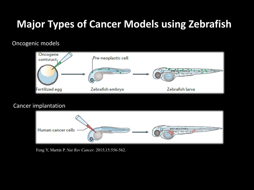
There are various methods for cancer research using zebrafish, but mainly two approaches are considered major.

1. Oncogenic models
By overexpressing oncogenic genes from humans or zebrafish in target organs, or by deleting tumor suppressor genes, zebrafish can be induced to develop cancer. Models that introduce the well-known BRAF (V600E) mutation gene associated with malignant melanoma into the skin of zebrafish, or liver cancer models, are commonly used.

2. Cancer implantation
This is a model where cancer cells derived from humans or other animals are transplanted into zebrafish. Success rates vary greatly depending on the transplantation site (yolk sac, duct of Cuvier, perivitelline space, intraperitoneal), stage of zebrafish (larvae, young, adult), and type of cancer cells used for transplantation, but there are numerous reports on this approach. 

Mobirise

Drug Screening using cancer xenograft zebrafish

We are conducting research on the screening of anti-cancer drugs that can eliminate transplanted cancer cells in zebrafish larvae, using cancer cells derived from various malignant tumors such as leukemia, malignant melanoma, colon cancer, breast cancer, etc. During this stage, zebrafish larvae are transparent, making it easy to quantify the amount of cancer cells in the body by expressing fluorescent proteins in the cancer cells. Cancer cell transplantation is performed through the duct of Cuvier, allowing for the study of distant metastasis by introducing cancer cells into the circulation. 

Mobirise

Zebrafish as a Tool for Anti-Cancer Drug Delivery Technology Development

Anti-cancer drugs generally have low specificity and can cause various side effects in normal tissues. To avoid these side effects, drug delivery technologies that specifically target cancer cells are essential. Cancer xenograft zebrafish, which have transparent bodies, allow monitoring of labeled cancer cells or anti-cancer drugs using fluorescent dyes or proteins, enabling confirmation of selective delivery of anti-cancer drugs to malignant tumors as shown in the figure. We have contributed to the development of various technologies, such as modified liposomes and micelles.

Mobirise
Address

2-174 Edobashi, Tsu, Mie, 5148572, Japan

Contacts

Yasuhito Shimada, MD, PhD.
Email: shimada.yasuhito@mie-u.ac.jp                     
Phone: +81 (0) 59 231 5384           

Easy Website Builder